
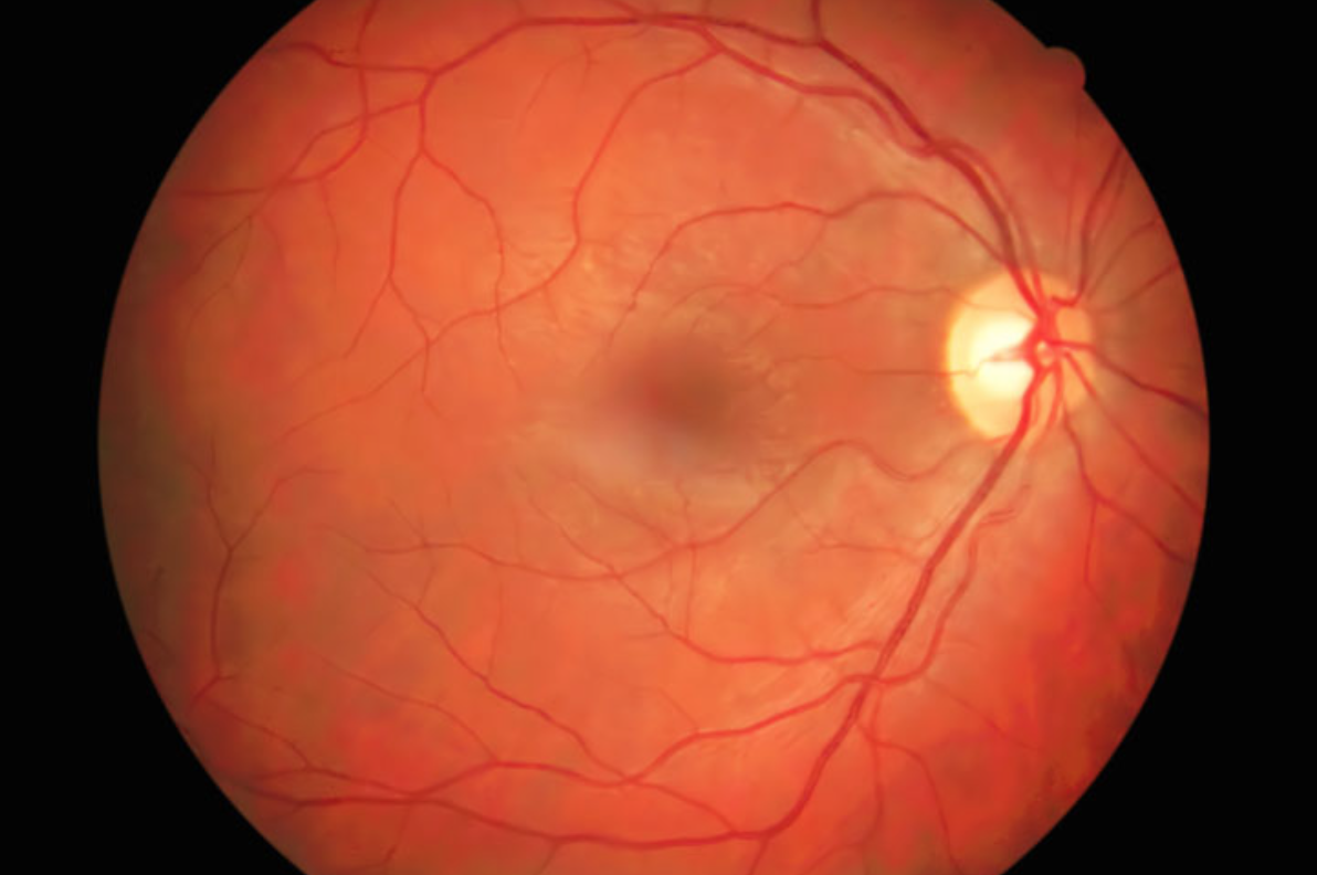
Retina specialists use fluorescein angiography (FA) to help diagnose and monitor a number of retinal diseases and conditions, including age-related macular degeneration, diabetic retinopathy, central serous retinopathy, macular edema, macular pucker, ocular melanoma/cancer, retinal artery occlusion (CRAO and BRAO) and retinal vein occlusion (CRVO and BRVO). It is a photographic study of the retina’s blood supply.
How It Works
Fluorescein angiography involves injecting a small amount of a yellow, vegetable-based dye through a patient’s peripheral vein – usually in the arm or hand. This dye travels through the blood vessels and lights up the retina’s detailed vascular network. A certified ophthalmic angiographer then takes a series of digital photographs of the eye using a special camera. These photographs help retina physicians identify and locate any leaking blood vessels, determine the amount of leakage, and detect any other abnormalities in the eye. The results allow them to develop a targeted treatment plan as well as track changes in eye disease over time.
Because it is vegetable-based, the dye used to perform an FA is generally safe and can be used on patients who have shellfish or iodine allergies as well as those who have previously experienced a negative reaction to contrast dye used in radiologic procedures. In very rare cases, a patient may experience an allergic reaction. Our staff will carefully monitor you throughout the test for any adverse reaction and provide appropriate treatment as necessary.
What to Expect
At Texas Retina Associates, fluorescein angiography (FA) is performed in our office and usually takes about 30 minutes.
- First, we will place drops in your eyes to dilate your pupils.
- Then the fluorescein dye is injected into a vein, usually in your arm or hand.
- It takes about 10-15 seconds for the dye to travel through your body to your eyes, and then the ophthalmic angiographer will digitally take the pictures using a specialized camera.
After the Fluorescein Angiography
- Immediately after the test, objects may appear dark or tinted, but this goes away within a few minutes.
- You should bring sunglasses to wear home as your eyes will be dilated.
- We also recommend that you have someone drive you home after your exam.
- Your urine may appear yellow, orange or green for a day or two after the test. This is normal and will clear up on its own.
- In addition, your skin may look slightly yellow for a few hours but will also return to normal.
Click here to learn more about what to expect from a visit to Texas Retina Associates.